


Description
ISBN 978-0-974-7694-55
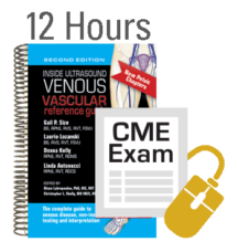
by: Gail P. Size BS, RPhS, RVS, RVT, FSVU,
Laurie Lozanski BS, RVT
Donna Kelly, RPhS, RVT, RDMS
Linda Antonucci, RPhS, RVT, RDCS
Updated and Expanded!
More Anatomy, Techniques, Cine Clips and New Pelvic and Ergonomics Chapters
Over 800 illustrations, images and videos, 55 tables and comprehensive information on:
- Upper Extremity Venous Anatomy
- Abdominal and Pelvic Veins Anatomy
- Lower Extremity Venous Anatomy
- Venous Vascular Physiology and Hemodynamics
- Venous Diseases
- ABI And Pedal Artery Waveforms
- Lower Extremity Venous Duplex Ultrasound
- Native Ivc, Iliac and Venous Stent Duplex
- Duplex Ultrasound for Pelvic Venous Disorders
- Lower Extremity Venous Insufficiency Duplex
- Venous Ablation Duplex
- Upper Extremity Venous Duplex
- Venous Photoplethysmography
- Air Plethysmography
- Ergonomics
- Correlative Testing Modalities
- Testing Optimization
- Cardiac Effects of PW Doppler
- Quality Improvement
- Measurements
- Glossary of Terms
- Acronyms
Full color images, anatomical drawings, quick reference tables and tips throughout the book, QR codes can be found throughout this book which will allow you to view video clips of various pathology or duplex findings using your smartphone or tablet.